Premium Only Content

DR CAMPRA PROVES GRAPHENE OXIDE IN COVID VACCINES
Distributed by: hackinthesystem.com | freedomwarrior.net
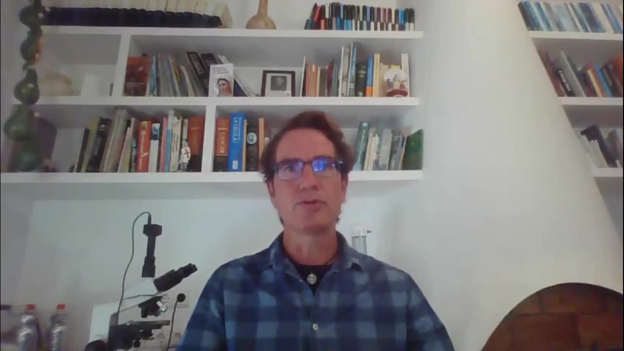
Dr Campra
Background
• Mr. Ricardo Delgado Martín requests PROVISION OF RESEARCH SERVICES to the UAL named:
"DETECTION OF GRAPHENE IN AQUEOUS SUSPENSION SAMPLE"
- On 06/10/2021 1 vial was received by courier, labeled with the following text:
- “COMIRNATY™ .Sterile concentrate. COVID-19 mRNA. 6 doses after dilution.
- Discard date / time: PAA165994.LOT / EXP: EY3014 08/2021 ”
- Origin and traceability: unknown
- State of conservation: refrigerated
- Maintenance during the study: refrigerated
- Coding of the problem sample to be analyzed: RD1
Preliminary observations of the test sample RD1
Description:
- Sealed vial, with rubber and aluminum cap intact, of 2 ml capacity, containing a 0.45 ml cloudy aqueous suspension.
- RNA extraction and quantification is performed
- Presence of uncharacterized nanometric microbiology is observed, visible at 600X in optical microscope
Sample processing
1. Dilution in 0.9% sterile physiological saline (0.45 ml + 1.2 ml)
2. Polarity fractionation: 1.2 ml hexane + 120 ul of RD1 sample
3. Extraction of hydrophilic phase
4. Extraction and quantification of RNA in the sample
5. Electron and optical microscopy of aqueous phase
Preliminary analysis: extraction and quantification of Rna in the sample
1. RNA extraction: Kit https://www.fishersci.es/shop/products/ambion-purelink-rna-mini-kit-7/10307963
2. Quantification of total UV absorbance in spectrophotometer
NanoDrop™ https://www.thermofisher.com/order/catalog/product/ND-2000#/ND-2000
3. Specific quantification of Rna by fluorescence QUBIT2.0:
UV absorption spectrum of the aqueous phase of the RD1 sample (Nano-drop team) Maximum absorption of SAMPLE RD1 (260-270 nm)
- RNA. It presents usual maximums at 260 nm. Total concentration estimated by QUBIT2.0 fluorometry: 6 ng / ul
- The spectrum reveals the presence of a high quantity of substances or substances other than Rna with maximum absorption in the same region, with a total estimated at 747 ng / ul (uncalibrated estimate)
- Reduced graphene oxide (RGO) has absorption maxima at 270 nm, compatible with the spectrum obtained (Thema et al, 2013. Journal of Chemistry ID 150536)
- The maximum absorption obtained DOES NOT ALLOW TO DISCARD the presence of graphene in the sample. The minimum amount of RNA detected by QUBIT2.0 only explains a residual percentage of the total UV absorption of the sample.
OBJECTIVE: Microscopic identification of graphene derivatives
METHODOLOGY:
1. Imaging in optical and electron microscopy
2. Comparison with literature images and reduced graphene oxide standard sample
TRANSMISSION ELECTRON MICROSCOPY (TEM)
Electron microscope JEM-2100Plus
Voltage: 200 kV
Resolution 0.14 nm
Magnification up to x1,200,000
TRANSMISSION ELECTRON MICROSCOPY (TEM)
Electron microscopy (TEM) is commonly used to image graphene nanomaterials. It has become a fairly standard and easy to use an instrument that is capable of imaging individual layered graphene sheets.
Download full report : https://hackinthesystem.com/dr-campra-detects-graphene-oxide-in-covid-19/
-
 1:49
1:49
HITSTV
2 years agoWEF - 5 Ways the scamdemic they deployed may change your life
137 -
 10:22
10:22
Dr Disrespect
2 days agoDR DISRESPECT - 2024 RECAP
17.2K69 -
 LIVE
LIVE
The Charlie Kirk Show
36 minutes agoThe 2025 Speaker Election + DEI FBI + Britain's Grooming Gangs | Gaetz, Gilliam, Marshall | 1.3.2025
9,663 watching -
 LIVE
LIVE
Grant Stinchfield
2 hours agoThe Left's Will Evil Plot to Prevent Trump from Taking Office Exposed!
659 watching -
 52:32
52:32
The Dan Bongino Show
3 hours agoProducer's Picks: Bongino's Best Segments - 01/03/25
60.9K252 -
 56:49
56:49
VSiNLive
2 hours agoA Numbers Game with Gill Alexander | Hour 1
6.31K1 -
 LIVE
LIVE
Film Threat
15 hours ago2025 MOVIES TO LOOK FORWARD TO + JANUARY'S BEST FILMS | Film Threat Livecast
268 watching -
 LIVE
LIVE
The Shannon Joy Show
3 hours ago🔥🔥Friday Freestyle LIVE With Shannon Joy! Ask Me Anything On The Open Live Chat!🔥🔥
290 watching -
 LIVE
LIVE
The Big Mig™
16 hours agoGlobal Finance Forum Powered By Genesis Gold Group
1,768 watching -
 1:31:22
Caleb Hammer
2 hours agoEgo-Maniac Thinks She Can Manifest Problems Away | Financial Audit
20.9K1