Premium Only Content

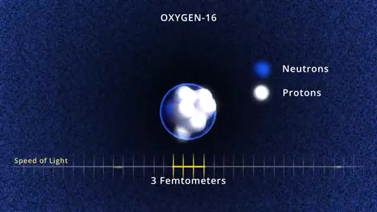
Visualizing the Nucleus
Visualizing the nucleus of a cell can provide a detailed understanding of its structure and function, which is critical for studies in cell biology, genetics, and medicine. Here are the most common approaches used to visualize the nucleus:
---
### **Light Microscopy**
1. **Fluorescence Microscopy**:
- **Dyes**: DNA-specific dyes like DAPI and Hoechst stain bind to DNA and emit fluorescence under UV light, highlighting the nucleus.
- **Fluorescent Proteins**: Genetically encoded fluorescent markers like GFP fused to nuclear proteins can illuminate the nucleus in live cells.
2. **Confocal Microscopy**:
- Offers high-resolution, 3D imaging of the nucleus by optically sectioning the sample.
3. **Phase Contrast and Differential Interference Contrast (DIC)**:
- These techniques provide non-stained visualization of the nucleus, though less detailed than fluorescence methods.
---
### **Electron Microscopy (EM)**
- **Transmission Electron Microscopy (TEM)**:
- Reveals ultrastructural details, including the nuclear envelope, nuclear pores, and chromatin distribution.
- **Scanning Electron Microscopy (SEM)**:
- Provides surface details of the nuclear envelope.
---
### **Advanced Imaging Techniques**
1. **Super-Resolution Microscopy**:
- Techniques like STED, PALM, or STORM break the diffraction limit, enabling visualization of nuclear features at nanometer resolution.
2. **Cryo-Electron Microscopy**:
- Captures high-resolution, near-native state images of the nucleus and its components.
---
### **Other Approaches**
1. **Live-Cell Imaging**:
- Time-lapse microscopy tracks nuclear dynamics in real-time.
2. **Atomic Force Microscopy (AFM)**:
- Measures topography and mechanical properties of the nucleus.
-
 LIVE
LIVE
Flyover Conservatives
21 hours agoUS STOCK MARKET: Sinking Ship - Dr. Kirk Elliott; How I Fought Back Against Woke Schools & Stopped Gender Bathrooms - Stacy Washington | FOC Show
830 watching -
 1:08:09
Donald Trump Jr.
8 hours agoFBI Dream Team, Plus Taking Your Questions Live! | Triggered Ep.219
135K216 -
 LIVE
LIVE
Akademiks
7 hours agoDrake and PartyNextDoor '$$$4U' Album Sells 250K first week. BIG AK IS BACK.
2,498 watching -
 3:12:08
MyronGainesX
6 hours ago $23.63 earnedDan Bongino Named As Deputy Director Of FBI And CPAC Recap
64.2K22 -
 3:12:31
vivafrei
6 hours agoBarnes Live from Seattle - Defending Benshoof in a Case that is CRAY CRAY!
78K26 -
 2:12:12
Robert Gouveia
6 hours agoLiberals EXPLODE over Elon's Email; Lawsuits FLY; Sanctions?? Congrats Dan!
48.3K25 -
 1:33:36
Redacted News
6 hours agoBREAKING! PUTIN LAUNCHES MASSIVE OFFENSIVE IN UKRAINE AS EUROPEAN LEADERS PUSH FOR MORE WAR
127K216 -
 44:39
44:39
Kimberly Guilfoyle
7 hours agoBetter Days Ahead for the FBI, Live with Asm Bill Essayli & John Koufos | Ep.199
82.3K25 -
 1:40:29
In The Litter Box w/ Jewels & Catturd
1 day agoWhat Did You Do Last Week? | In the Litter Box w/ Jewels & Catturd – Ep. 748 – 2/24/2025
127K36 -
 23:34
23:34
Stephen Gardner
7 hours ago🔥CNN PANICS over $5000 DOGE Dividend | Trump Orders bigger Audits
77.3K132