Premium Only Content

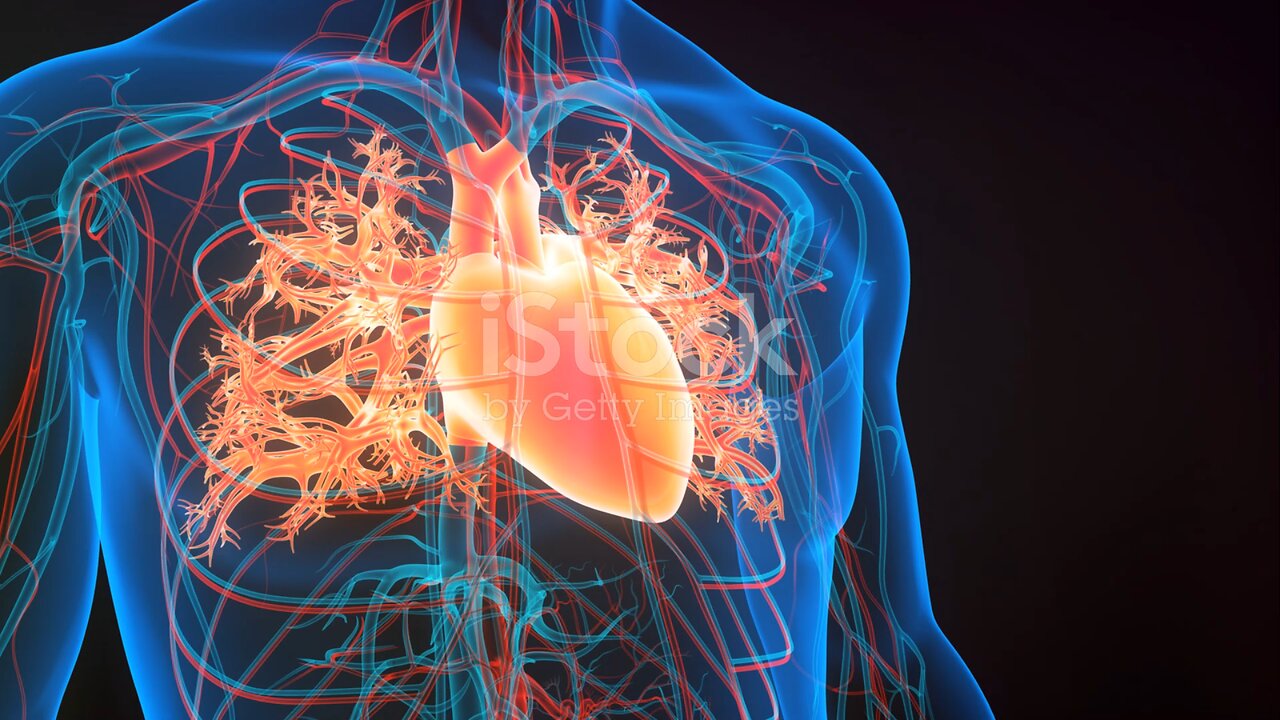
Humen heart anatomy (Basic)
Certainly! Here's a more detailed description of the anatomy of the human heart:
1. **Pericardium:** The heart is surrounded by a double-walled sac called the pericardium. The outer layer is called the fibrous pericardium, which is tough and protective. The inner layer is the serous pericardium, consisting of two layers: the parietal layer (lining the fibrous pericardium) and the visceral layer (also known as the epicardium, which covers the heart muscle).
2. **Heart Wall:** The heart wall is composed of three layers:
- **Epicardium:** The outermost layer, as mentioned, is the visceral layer of the serous pericardium.
- **Myocardium:** The middle layer, consisting of cardiac muscle tissue responsible for the pumping action of the heart.
- **Endocardium:** The innermost layer, a thin layer of endothelial tissue that lines the chambers of the heart and covers the heart valves.
3. **Chambers:** The heart has four chambers:
- **Right Atrium:** Receives deoxygenated blood returning from the body via the superior and inferior vena cava.
- **Right Ventricle:** Receives blood from the right atrium and pumps it to the lungs for oxygenation through the pulmonary artery.
- **Left Atrium:** Receives oxygenated blood from the lungs via the pulmonary veins.
- **Left Ventricle:** Receives blood from the left atrium and pumps it to the body through the aorta.
4. **Valves:** Valves regulate blood flow between the chambers of the heart and prevent backflow:
- **Tricuspid Valve:** Located between the right atrium and right ventricle.
- **Pulmonary Valve:** Positioned between the right ventricle and pulmonary artery.
- **Mitral Valve (Bicuspid Valve):** Found between the left atrium and left ventricle.
- **Aortic Valve:** Located between the left ventricle and the aorta.
5. **Blood Vessels:**
- **Coronary Arteries:** Supply oxygenated blood to the heart muscle.
- **Coronary Veins:** Drain deoxygenated blood from the heart muscle.
- **Pulmonary Arteries and Veins:** Transport blood between the heart and lungs for oxygenation.
6. **Electrical Conduction System:** The heart has its own electrical conduction system, including the sinoatrial (SA) node, atrioventricular (AV) node, bundle of His, and Purkinje fibers. This system controls the heart rate and rhythm.
Understanding the detailed anatomy of the heart is crucial for comprehending its function and the pathophysiology of various cardiac conditions.
-
 4:53:35
Rotella Games
9 hours agoGrand Theft America - GTA IV | Day 4
42K4 -
 LIVE
LIVE
Scottish Viking Gaming
7 hours ago💚Rumble :|: Sunday Funday :|: Rumble Fam Knows What's Up!!
274 watching -
 LIVE
LIVE
ttvglamourx
6 hours ago $2.69 earnedEGIRL VS TOXIC COD LOBBIES !DISCORD
305 watching -
 3:19:17
LumpyPotatoX2
8 hours agoSCUM: Lumpy Land RP Server - Day #1 - #RumbleGaming
57.9K2 -
 1:42:59
Game On!
21 hours ago $8.68 earnedTop 10 Super Bowl Bets You Can't Afford To Miss!
87K9 -
 2:17:02
Tundra Tactical
1 day ago $22.75 earnedTundra Nation Live : Shawn Of S2 Armament Joins The Boys
170K28 -
 11:00:11
tacetmort3m
1 day ago🔴 LIVE - SOLO RANK GRINDING CONTINUES - MARVEL RIVALS
215K5 -
![Shadows Of Chroma Tower, Alpha Playtest [Part 1]](https://1a-1791.com/video/fwe2/1d/s8/1/5/Q/U/n/5QUnx.0kob-small-Shadows-Of-Chroma-Tower-Alp.jpg) 13:29:21
13:29:21
iViperKing
1 day agoShadows Of Chroma Tower, Alpha Playtest [Part 1]
176K9 -
 54:05
54:05
TheGetCanceledPodcast
1 day ago $13.90 earnedThe GCP Ep.11 | Smack White Talks Smack DVD Vs WorldStar, Battle Rap, Universal Hood Pass & More...
150K35 -
 13:37
13:37
Exploring With Nug
1 day ago $10.52 earnedSUV Found Underwater Searching For Missing Man Jerry Wilkins!
108K5