Premium Only Content

The Late Prof. Arne Burkhardt - Autopsies: Evidence for Jab Related Harm and Death. Feb. 5, 2022
The inaugural Understanding Vaccine Causation Conference, convened by World Council for Health- Prof. Arne Burkhardt full demonstration: https://rumble.com/v3yajuy-prof.-arne-burkhardt-autopsies-evidence-for-jab-related-harm-and-death-feb-.html?mref=1bxo9j&mrefc=20
The Late Prof. Arne Burkhardt joined the Medical Practice panel to share his presentation, Autopsies: Evidence for Jab Related Harm and Death. Feb. 5, 2022
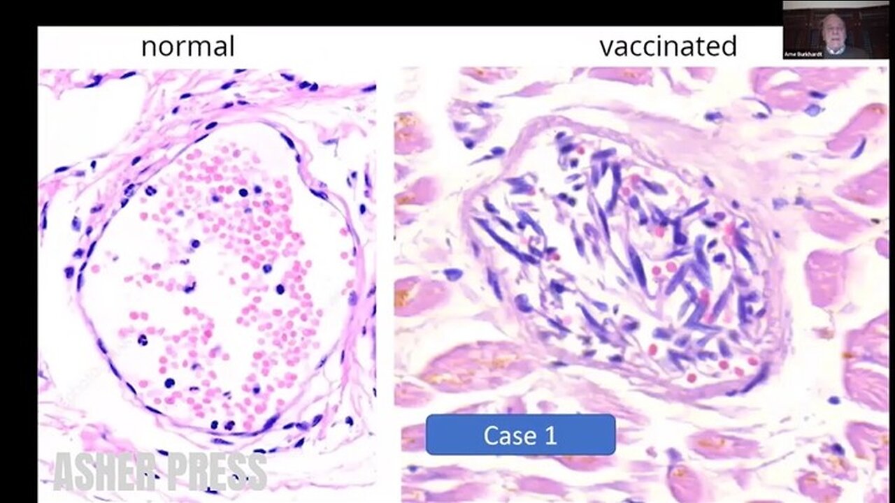
(Transcript) And I will just show you a few examples of the tissue damage that I have listed before. So here you can see a normal, small vessel, and you can see the Endothelium that is like a wallpaper and very small elongated spindle cell nuclei. And here in one of the cases, you can see that the Endothelium is in the lumen. And it is in there mixed with Lymphocytes and Erythrocytes and the nuclei is swollen.
So in some cases the small vessels even are completely destroyed by inflammatory infiltrates, mostly Lymphocytes. And this proves to me that it is an intravital reaction and not an autorhythmic phenomenon caused by degradation after death.
So we get the spike protein, immunohistochemistry on these cases and we see, you see here a very marked and specific, eh, the mark of the endothelium in these patients. And not only in the small vessels, but also in the smaller arteries, you can see it in the inner part of the vessel. And you can see here there’s decimated endothelial cells.
So not as I said, not only the smaller vessels were affected, but also the aorta and the larger arteries and two cases have died of a ruptured artery. And actually we found arteriosclerotic changes, but as you see here, it’s not very pronounced. But you can see inflammatory changes around in the deep layers of this aorta and also you can see some disturbance of structure of the smooth muscle and the elastic fibers. And if you have a higher magnification, you can see these small areas where the elastic fibers and smooth muscles are destroyed.
And again, lymphocytic infiltration proving that it was an intravital process. And here another case. We found it in, in five cases so this cannot be a coincidence. And we did the spike protein, and you find a marked positive expermeation of spike protein in the myofibroblasts of the arterial wall. This is the aorta. And also in the [unintelligible], you can see very strong expression of spike protein in these areas. And this I think is a very important finding.
So how often did we see this vasculitis, this endovasculitis or some call it endothelitis, in 11 cases with focal lymphocytic infiltration, then vasculitis paravasculitis in 10 cases, focal media-necrosis in six cases, and thrombosis caused on this area in two cases.
https://worldcouncilforhealth.org/multimedia/uvc-arne-burkhardt/
-
 0:11
0:11
Asher Press
1 day agoThat's all folks!
3601 -
 1:49:14
Redacted News
5 hours agoTrump is Back! Congress Uncovers New Biden Crimes One Day After He Leaves D.C. | Redacted
126K193 -
 2:09:53
Benny Johnson
5 hours ago🚨President Trump LIVE Right Now Making MASSIVE Announcement At White House News Conference
215K235 -
 2:04:10
Revenge of the Cis
6 hours agoEpisode 1433: Retribution
82.9K13 -
 1:42:50
The Criminal Connection Podcast
10 hours ago $0.57 earnedEddie Hearn talks JOSHUA vs FURY, Working With Frank Warren & The Truth About Turki Alalshikh!
32.4K2 -
 1:00:25
In The Litter Box w/ Jewels & Catturd
1 day agoGolden Age | In the Litter Box w/ Jewels & Catturd – Ep. 724 – 1/21/2025
125K57 -
 57:42
57:42
The Dan Bongino Show
13 hours agoHE'S BACK! (Ep. 2405) - 01/21/2025
1.32M2.17K -
 46:19
46:19
Candace Show Podcast
7 hours agoUH-OH! Elon’s Viral Salute Steals The Inauguration Show | Candace Ep 136
136K363 -
 8:05:01
hambinooo
11 hours agoNO COMMIE TUESDAY
90.3K3 -
 2:08:37
The Quartering
8 hours agoTrump Ends Online Censorship, Foreign Aid, Frees J6 Hostages & Much More In Day 1
130K46