Premium Only Content

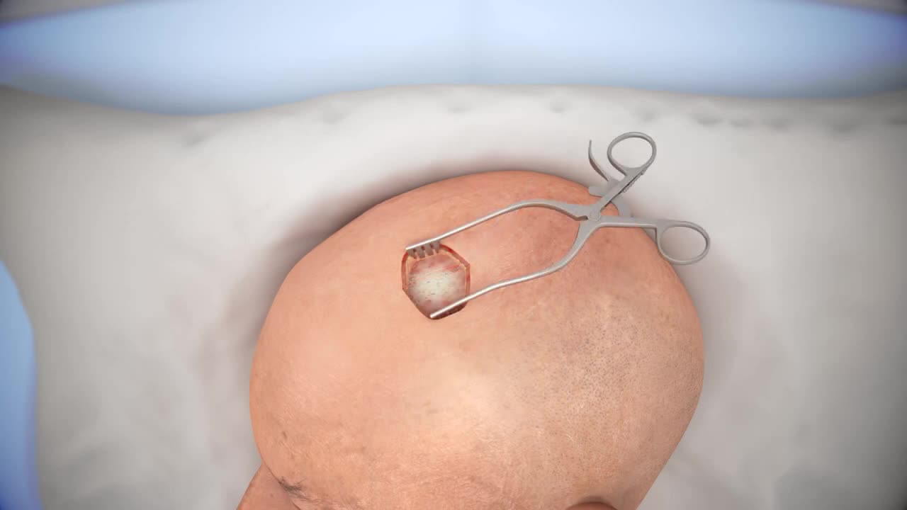
Ventriculostomy Brain Surgery - 3d animation
This 3D animation of brain surgery, shows how a ventriculostomy is performed, which is a neurosurgical procedure of creating a hole within a cerebral ventricle for drainage. It is most commonly performed on those with hydrocephalus, an abnormal buildup of fluid in the ventricles (cavities) deep within the brain. It's done by surgically penetrating the skull, dura mater, and brain such that the ventricular system ventricle of the brain is accessed.
When catheter drainage is temporary, it is commonly referred to as an external ventricular drain (EVD). When catheter drainage is permanent, it is usually referred to as a shunt.
There are many catheter-based ventricular shunts that are named for where they terminate, for example, a ventriculi-peritoneal shunt terminates in the peritoneal cavity, a ventriculoarterial shunt terminates within the atrium of the heart, etc. The most common entry point on the skull is called Kocher's point. An EVD ventriculostomy is done primarily to monitor the intracranial pressure as well as to drain cerebrospinal fluid (CSF), primarily, or blood to relieve pressure from the central nervous system (CNS).
#Pharmaceutical #Biotechnology #Therapeutics #Marketing #Training #Education #3DAnimation #Medicine #Medical Sciences #Surgery #MOA Medicine #Drugs #Anatomy #Technology #Medical Animation #3DAnimation #Mechanism Of Action #Surgical Procedure #Surgical Video #HealthCare #Medical Animation Companies #Important Videos #Medical Devices #Surgical Procedures #3DVideo #3D Medical Animation #Medical Procedure #Amerra Medical #Nucleus Medical #Scientific Animation
-
 1:19:48
Simply Bitcoin
12 hours ago $6.75 earnedJerome Powells MASSIVE Bitcoin Backflip! | EP 1172
56.9K5 -
 58:42
58:42
The StoneZONE with Roger Stone
4 hours agoLBJ + CIA + Mob + Texas Oil = JFK Murder | The StoneZONE w/ Roger Stone
41.7K24 -
 58:00
58:00
Donald Trump Jr.
11 hours agoBreaking News on Deadly Plane Crash, Plus Hearing on the Hill, Live with Rep Cory Mills & Sen Marsha Blackburn | TRIGGERED Ep.212
174K135 -
 52:03
52:03
Kimberly Guilfoyle
10 hours agoLatest Updates on Deadly Air Collision, Plus Major Hearings on Capitol Hill,Live with Marc Beckman & Steve Friend | Ep.192
98.2K37 -
 1:17:16
Josh Pate's College Football Show
8 hours ago $1.40 earnedMichigan vs NCAA | ESPN’s ACC Deal | Season Grades: UGA & Miami | Notre Dame Losses
39.1K2 -
 1:26:50
Redacted News
8 hours agoWhat happened? Trump DESTROYS the Pete Buttigieg run FAA for tragic airline crash | Redacted News
225K185 -
 LIVE
LIVE
VOPUSARADIO
1 day agoPOLITI-SHOCK! Hero Angela Stanton King & Wrongfully Imprisoned J6 Prisoner Josh Pruitt!
74 watching -
 43:37
43:37
Candace Show Podcast
8 hours agoThe Taylor Swift Plot Thickens | Candace Ep 142
144K126 -
 1:37:33
Common Threads
6 hours agoLIVE DEBATE: Trump's Wild Handling of Plane Tragedy
27.1K8 -
 54:45
54:45
LFA TV
13 hours agoConfirmation Chaos | TRUMPET DAILY 1.30.25 7pm
38.5K5