Premium Only Content

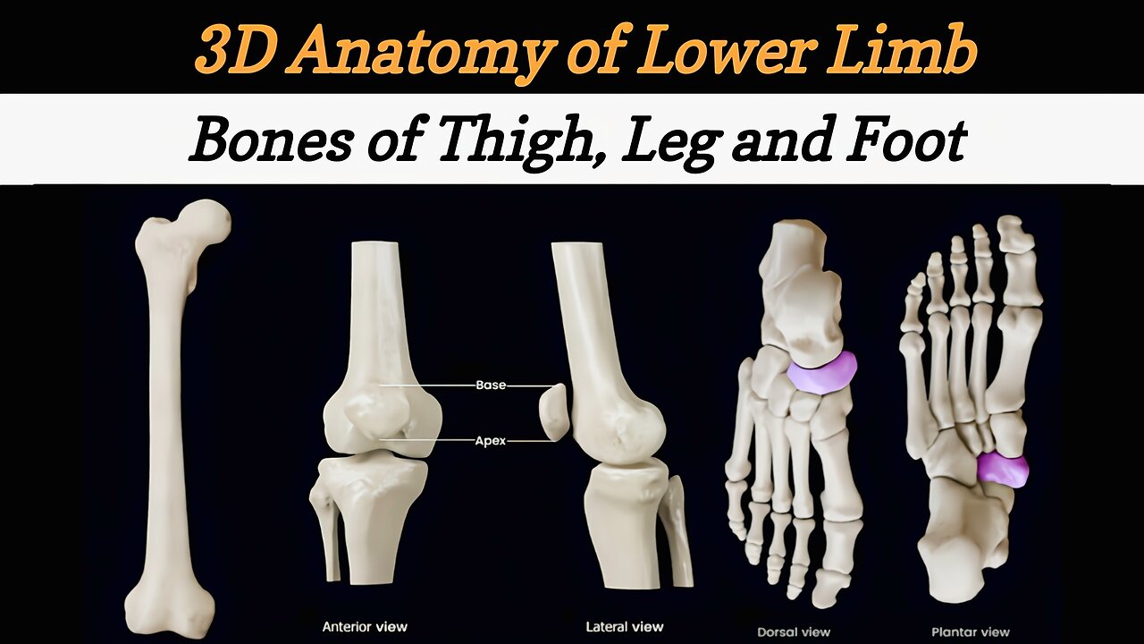
Lower Limb Bones | 3D Anatomy of Lower Limb | Revision of Lower Limb Bones | Anatomy - Lecture #5
Bones of Lower Limb | How to Remember every bone of Lower Limb | Hip Bone and femur | Tibia and Fibula | Femur Bone Anatomy 3D | Bones and Joints | Foot Bones | निचले अंग की शारीरिक रचना | nichale ang kee shaareerik rachana
Lower limb anatomy refers to the study of the structures and components of the lower extremities of the human body. The lower limb, also known as the lower extremity, consists of the hip, thigh, knee, leg, and foot. Here are the main structures and bones of each region:
Hip:
Hip joint: The ball-and-socket joint formed by the femur (thigh bone) and the acetabulum of the pelvis.
Thigh:
Femur: The longest and strongest bone in the body, located between the hip and the knee.
Patella (Kneecap): A small bone in front of the knee joint.
Knee:
Knee joint: A hinge joint formed by the femur, tibia (shinbone), and patella.
Tibia: Also known as the shinbone, it is the larger bone of the lower leg.
Fibula: A slender bone located alongside the tibia in the lower leg.
Leg:
Anterior Compartment:
Tibialis anterior: Located on the front of the leg, it helps with dorsiflexion and inversion of the foot.
Extensor digitorum longus: Extends the toes and dorsiflexes the foot.
Extensor hallucis longus: Extends the big toe and dorsiflexes the foot.
Posterior Compartment:
Gastrocnemius: The calf muscle that flexes the knee and plantarflexes the foot.
Soleus: Located beneath the gastrocnemius, it also helps with plantarflexion.
Tibialis posterior: Located deep within the leg, it helps with inversion and plantarflexion of the foot.
Medial Compartment:
Flexor digitorum longus: Flexes the toes.
Flexor hallucis longus: Flexes the big toe.
Tibialis posterior: Also present in the medial compartment, it helps with inversion and plantarflexion.
Foot:
Tarsal bones: Seven bones that make up the posterior part of the foot, including the calcaneus (heel bone), talus, navicular, cuboid, and three cuneiform bones.
Metatarsal bones: Five long bones located in the middle of the foot, connecting the tarsal bones to the phalanges.
Phalanges: The bones of the toes, with three phalanges in each toe (except the big toe, which has two).
______________________________________________
Introduction of Anatomy Lecture #1
https://youtu.be/6jlKiz8FDdA
Anatomical position and directional terms Lecture #2
https://youtube.com/shorts/onwhUzFBBzk?feature=share
Bones of the Skull Lecture #3
https://youtu.be/qeouXzYBYKo
Bones of Upper Limb Lecture #4
https://youtu.be/f17CbBbQox8
______________________________________________
For Queries: mshabiulhasnain781@gmail.com
Show your support here
INSTAGRAM : @learnwithdrshabi
Link : https://instagram.com/learnwithdrshabi?igshid=NGExMmI2YTkyZg==
TIKTOK : @shabi1272
Link : http://tiktok.com/@learnwithdrshabi
#anatomy #lowerlimb #bones #learnwithDrShabi #bonesoflowerlimb #education #drnajeeb #drjayapaul #lecture #medical #medicalstudent #angelinaissac #pharmacy #pharmacytechnician @SamWebster @armandohasudungan @Corporis @AngelinaIssac @Kenhub @JohariMBBS @professorfink #lowerlimbanatomy #femur #tibia #fibula #foot #hipjoint #kneejoint
-
 18:54
18:54
The Rubin Report
52 minutes agoHow One Woman Outsmarted Pornhub & Exposed Its Dark Secrets | Laila Mickelwait
1 -
 LIVE
LIVE
Major League Fishing
4 days agoLIVE! - Bass Pro Tour: Stage 3 - Day 4
1,280 watching -
 1:05:28
Sports Wars
3 hours agoLebron GOES OFF Over Bronny Hate, Pereira LOSES Belt To Ankalaev At UFC 313, Xavier Worthy Arrested
3.85K4 -
 10:27
10:27
Tactical Advisor
1 day agoDMR or SPR for Civilian Use?
25.3K5 -
 8:21
8:21
DEADBUGsays
1 day agoThe Crossbow Killer
12.1K8 -
 8:40
8:40
Tundra Tactical
20 hours ago $5.53 earnedThe Executive Order Wishlist.
25.1K2 -
 7:22:52
SpartakusLIVE
19 hours agoSaturday SPARTOON Solos to Start || Duos w/ StevieT Later
104K2 -
 28:40
28:40
SLS - Street League Skateboarding
8 days agoTOP MOMENTS IN WOMEN’S SLS HISTORY! ALL THE 9’s - Rayssa Leal, Leticia Bufoni, Chloe Covell & more…
71.8K10 -
 2:03:03
The Connect: With Johnny Mitchell
17 hours ago $3.88 earnedHow Mexican & Chinese Cartels Control Illegal Marijuana Cultivation In America Using SLAVE Labor
20.2K4 -
 14:46
14:46
Mrgunsngear
18 hours ago $2.21 earnedPrimary Arms GLx 1x Prism With ACSS Reticle Review
26K8