Premium Only Content

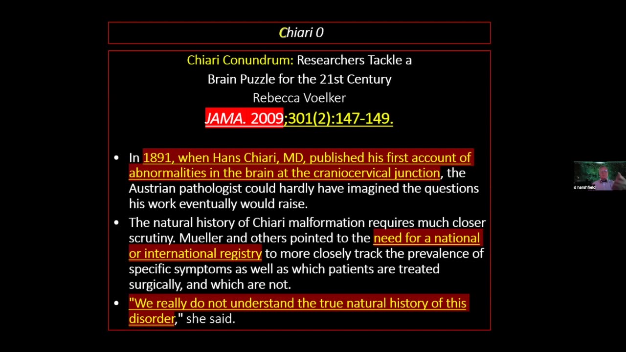
53. Craniocervical Junction Abnormalities (only) Dr. Harshfield
Resources from video chat:
12:42:22 From Doug Leonard : Craniosacral osteopaths know this and treat the junction all the time! Quess what I do
12:45:57 From Susan : what kind of adjustment was that?
12:46:32 From Russ Allen : Atlas Orthogonist
12:46:33 From Dr. Tom Lewis : C1 LOW VELOCITY ATLAS
Doug, is there an association which shows us how to locate local Craniosacral osteopaths?
12:51:04 From - : https://nucca.org/
12:51:09 From Dr. Tom Lewis : SWEAT INSTITUTE IN ATLANTA
12:51:23 From Louise Vogel : I am currently having my C1 repositioned by a chiropractor in PA, Marc Calicchio who trained w Dr. H’s colleague Dr. Scott Rosa. He doesn’t have the upright MRI and instead does x-rays which he interpolates from.
12:51:48 From - : http://www.sweatinstitute.com/t2/index.htm - Welcome To The Sweat Institute
12:51:52 From integrative brain : Are you utilizing Dr Rugierio's work on lymph drainage w/ Dr. D. Klinghardt. He has lymph cream and massage to open the pathway for brain drainage?
12:52:22 From Steve : Replying to "http://www.sweatinst..."
Thanx!
12:52:23 From - : https://nucca.org/ - Welcome to National Upper Cervical Chiropractic Association
12:52:58 From Doug Leonard : Replying to "Craniosacral osteopa..."
https://cranialacademy.org/
12:56:10 From - : What to Expect From Atlas Orthogonal Technique
- https://uppercervicalawareness.com/what-to-expect-from-atlas-orthogonal-technique/
13:01:23 From - : https://www.hudsonvalleyscoliosis.com/treatments/khan-kinetic-treatment/
13:03:00 From - : https://www.kktspine.com/kktstory/# - THE KKT TREATMENT PROCESS
13:03:16 From Dr. Tom Lewis : To get to Dr. Harshfield: akfahoum@gmail.com
Craniocervical junction abnormalities are congenital or acquired abnormalities of the occipital bone, foramen magnum, or first two cervical vertebrae that decrease the space for the lower brain stem and cervical cord. These abnormalities can result in neck pain; syringomyelia; cerebellar, lower cranial nerve, and spinal cord deficits; and vertebrobasilar ischemia. Diagnosis involves magnetic resonance imaging (MRI) or computed tomography (CT).
-
 1:27:50
HEALTH EXPOSED By Health Revival Partners
2 months agoCancer is MAINLY a disease of Immune Deficiency - Dr. Lewis
466 -
 2:25:43
Darkhorse Podcast
12 hours agoLooking Back and Looking Forward: The 258 Evolutionary Lens with Bret Weinstein and Heather Heying
132K203 -
 5:50:16
Pepkilla
11 hours agoRanked Warzone ~ Are we getting to platinum today or waaa
90.2K7 -
 DVR
DVR
BrancoFXDC
9 hours ago $6.29 earnedHAPPY NEW YEARS - Road to Platinum - Ranked Warzone
81.3K2 -
 5:53
5:53
SLS - Street League Skateboarding
5 days agoBraden Hoban’s San Diego Roots & Hometown Win | Kona Big Wave “Beyond The Ride” Part 2
91.1K13 -
 6:03:57
TheBedBug
13 hours ago🔴 LIVE: EPIC CROSSOVER - PATH OF EXILE 2 x MARVEL RIVALS
94.5K9 -
 1:12:45
The Quartering
11 hours agoTerror In New Orleans, Attacker Unmasked, Tesla BLOWS UP At Trump Tower! Are We Under Attack?
157K254 -
 1:32:08
Robert Gouveia
13 hours agoNew Year TERROR; Trump Speaks at Mar-a-Lago; Speaker Johnson FIGHT
128K107 -
 22:21
22:21
Russell Brand
1 day agoVaccines Don't Cause Autism*
198K791 -
 2:05:27
The Dilley Show
13 hours ago $25.56 earnedNew Years Agenda, New Orleans Terror Attack and More! w/Author Brenden Dilley 01/01/2025
116K39