Premium Only Content

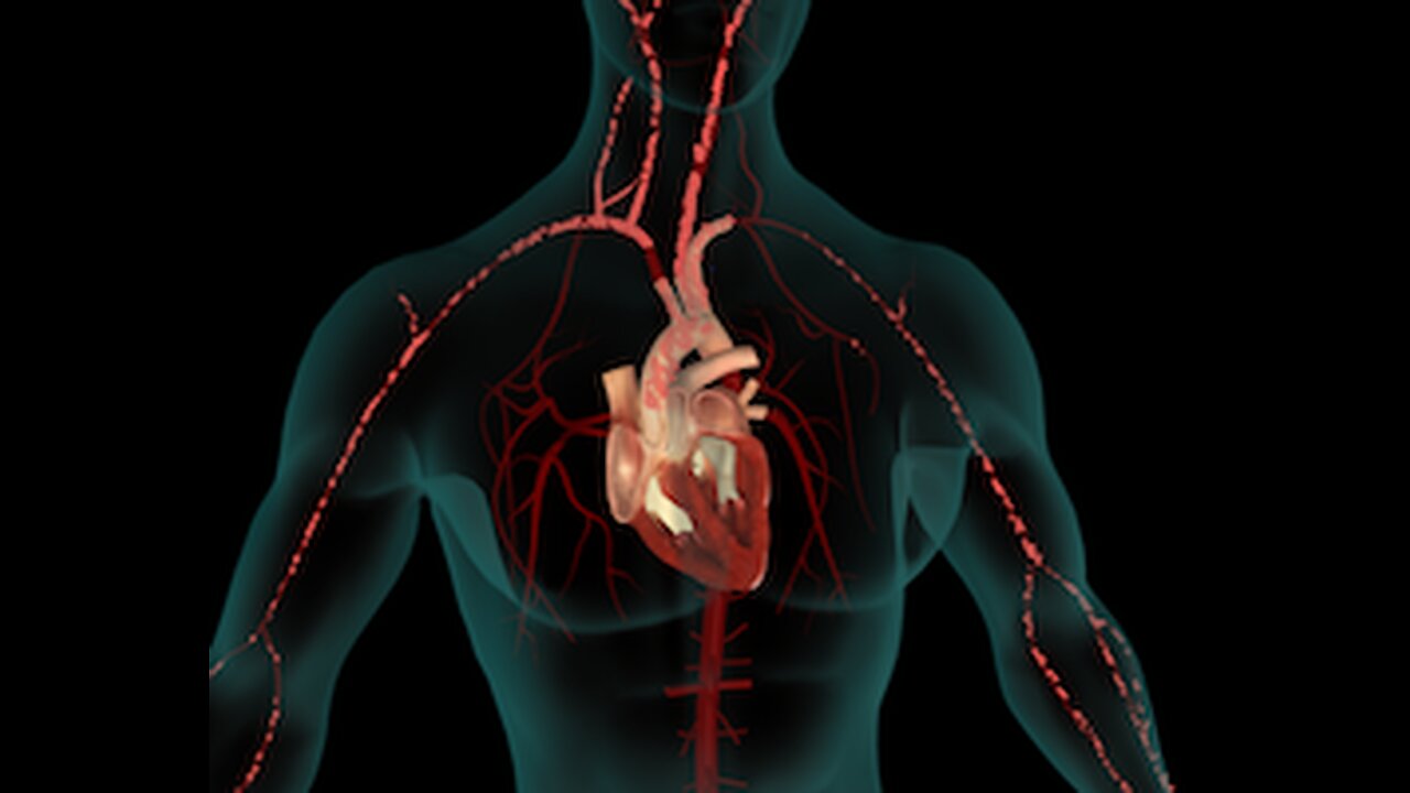
An In-Depth Look at the Circulatory System
Full video about Circulatory System
===
[00:00:00] The circulatory system is responsible for transporting oxygen-rich blood, or oxygenated blood and nutrients throughout the body and removes carbon dioxide. It consists of a massive muscular heart and a system of closed blood vessels comprising arteries. Arterials capillaries is veles and veins, and hence is also known as the cardiovascular.
It acts as the connecting link between the neuromuscular system and organs, such as the digestive, respiratory, excretory, organs, et cetera. The heart transports blood to the tissues to maintain their normal functioning. The blood collects oxygen from the lungs and carries it to the tissues and cells while removing their gaseous waste as carbon dioxide and directing it back to the lung.
The blood absorbs nutrients such as glucose, amino acids, and fatty [00:01:00] acids from the digestive system, and carries these energy rich tissue building products to all the cells of the body. It carries the waste products of cellular metabolism to the kidneys and other excretory organs. It also helps to coordinate the activities of various organs by transporting the hormones to the target organ.
There are two circuits of circulation, and both these circuits start from the heart and terminate in the heart. The blood is kept under circulation by two pumps of the heart, the right sided consisting of the right atrium and right ventricle, and the left sided consisting of the left atrium and left ventricle.
This kind of circulation is called double circulation. The two circuits of circulation are the systemic circulation and the pulmonary circulation. The coronary circulation, which supplies blood vessels to the muscular wall of the heart may be considered as a separate circuit. [00:02:00] Systemic circulation refers to pumping of oxygenated blood throughout the body from the left ventricle, and bringing back the oxygenated blood from the body to the right atr.
The oxygenated blood from the left ventricle is pumped into the great aorta, which arches itself to the left side and runs down as the descending aorta. The descending aorta through a system of arteries, supplies blood to various parts of the body where they end up in capillaris. The exchange of materials, mainly nutrients and oxygen, takes place between the wall of the capillaries and the tissue.
Waste products produced by the tissue cells such as carbon dioxide and urea are collected by capillaries that unite to form veles. The veles unite into larger vessels called veins. These veins again unite to form the superior vena cava and the inferior [00:03:00] vena cava, which bring the deoxygenated blood to the right atrium from where it flows into the right ventric.
Pulmonary circulation refers to pumping of deoxygenated blood from the right atrium to the lungs for purification and bringing back oxygenated blood from the lungs to the left atrium of the heart. The deoxygenated blood from the right ventricle is pumped into the lungs through pulmonary arteries in the lungs.
The blood passes through the alveola. Cappi is where gas exchange takes place removing carbon dioxide from blood cells. And enriching them with oxygen from the lungs. The oxygenated blood is then taken back to the left atrium by two pulmonary veins from each lung, thus four pulmonary veins empty oxygenated blood into the left atrium from where blood is further pumped into circulation.
The great aorta immediately after its [00:04:00] origin gives rise to the coronary arteries, which supply oxygenated blood and nutrients to the walls of the heart. There are two coronary arteries, the left coronary artery, L C A, and the right coronary artery, RCA A. The left coronary artery divides into two main branches.
One branch descends down as left anterior descending artery, L a. Which gives one or more branches called diagonals D. The second branch is called the circumflex, C I R C, which goes along the left margin of the heart and branches off into obtuse marginals. Om blood is drained from the heart muscles by a large vein, which terminates in a large venous trunk, the coronary sinus, which subsequently drains the blood from the wall of the heart to the right atrium.
The heart is a fist sized, cone-shaped, massive muscular [00:05:00] organ protected in the chest cavity. The pointed end of the cone called the apex of the heart is directed downwards and tilted a little to the left side. The wall of the heart is composed of three layers, an outer thin layer called Epicardium, the middle muscular layer called myocardium.
And the innermost thin and smooth lining called the endocardium. The heart is covered on the outer side by a double layered sack called the pericardium. The space between the pericardium and the epicardium is filled with a protective fluid called pericardial fluid. The myocardium is composed of a characteristic muscle called cardiac muscle.
Which alternatively contracts and relaxes in a regular clock like manner throughout life, the heart is divided into four chambers, namely the right and left atria, oracles, and [00:06:00] the right and left ventricles. The septum between the thin walled oracles is called inter septum, and the one between the thick walled ventricles is called inter ventricular Sept.
The right atrio. Ventricular aperture is provided with the tricuspid valve and the left atrio. Ventricular aperture is guarded by the bicuspid valve. The opening of the pulmonary artery into the right ventricle is guarded by the semilunar valve or pulmonary valve, and the opening of the aorta into the left ventricle is guarded by the aortic valve, which is also known as semilunar valve.
The right side of the heart pumps, deoxygenated blood or oxygen, poor blood, and the left side pumps oxygenated blood or oxygen-rich blood. The valves of the heart prevent backflow of blood into the chambers. The blood is kept circulating by the two pumps of the heart. One pump [00:07:00] pulmonary pump, represented by the right atrium and right ventric.
Receives deoxygenated blood from the systemic circulation and passes it onto the lungs. The second pump, systemic pump, represented by the left atrium and left ventricle, receives oxygenated blood from the pulmonary circulation and passes it on to all parts of the body for supplying oxygen to the cells.
The circulation of blood from the heart through pulmonary and systemic circulation and back to the heart again, is kept going by the rhythmical activity of the cardiac muscle. The phase of contraction is called sisterly, and the phase of relaxation is called diastole. The alternate contraction and relaxation of the oracles and the ventricles, followed by a short pause, is called the cardiac cycle, which constitutes the heartbeat.
The normal heart beats 60 to 80 times per minute, pumping five to six [00:08:00] liters of blood in a minute. Mild exercise raises the heartbeat to 100 to 120 times a minute, whereas strenuous exercise increases the heartbeat to up to 200 times a minute, pumping almost 30 liters of blood per minute. The blood is a fluid connective tissue, which is composed of plasma, different types of cells called cortisols and platelet.
Blood plasma is about 90% water, and the other 10% is protein, fat, glucose, and waste materials. Blood also contains clotting factors essential for coagulation of blood. The plasma from which clotting factors have been removed is called serum. The formation of new blood cells takes place in the bone marrow.
These blood cells then mobilize to blood circula. RBCs are circular, dislike by concave cells without nuclei. RBCs are red [00:09:00] in color due to the presence of red respiratory pigment hemoglobin, which is responsible for transporting oxygen two and carbon dioxide away from all parts of the body. The lifespan of RBCs is of 100 to 125 days.
They are then filtered outta the bloodstream by the spleen or the. The WBCs are devoid of hemoglobin and hence are colorless. WBCs live from six to nine days and are then absorbed by the spleen. WBCs defend the body by destroying pathogens, either by their phagocytic activities or by forming antibodies.
These are mainly of two types, the granulocytes or polymorph, nuclear leukocytes, and a granulary. Granulocytes are those with granules in the cytoplasm, and these include neutrophils, eosinophils and basophils. [00:10:00] A granulocytes are without granules in the cytoplasm. These include lymphocytes and monocytes.
The blood platelets are fragmented bodies without nuclei. These play an important role in blood coagulation. Blood grouping is the system of classification of blood according to different antigens on the surface of the red blood cells and the antibodies in the blood. The two main blood grouping systems are a b O system and rhus system.
Blood serum contains two antibodies. A glutenins and RBCs contain two antigens. A glutino gins antigens are designated as a and. And the corresponding antibodies are designated as antia or alpha and anti B or beta. An individual's blood group is denoted by the type of antigen present on RBCs. A glutenins cause clumping, [00:11:00] agglutination of the blood cells of individuals of the same species.
On this basis, the reactions of serum and RBCs fall into four groups a. A, B and O. The blood of persons belonging to group A contains a antigen and anti B antibody. Blood of B group contains B antigen and anti A antibody. Persons bearing the a b blood group have both A and B antigens, but no antia or anti B antibodies.
People with O Blood group do not contain any antigens, but they contain both antia and anti B antibodies in blood transfusions. It is always desirable to use a donor of the same group as the recipient to prevent clumping of RBCs. Rhus factor are H Factor is an antigen present on the surface of red blood.
Depending on the [00:12:00] presence or absence of this antigen, an individual is grouped as rhus positive r h positive and RHUS negative are H negative. Apart from being important in grouping the blood during blood transfusion, the RH system is very much necessary to determine the risk of hemolytic disease of the newborn, also known as Erythroblastosis, Fatali Group A.
Parents are of two types. Namely those with Group A progeny and those with group A and group O progeny. Similarly, group B parents are of two types, namely those with group B progeny and those with B and O progeny. The progeny of a B parents may be A B or A B. The progeny of O parents will be O only.
Genetically A, B, a, B and O. Blood groups are determined and controlled by a series of multiple [00:13:00] alleles. The gene is designated as I, since the antigens involved, unknown as ISO or glutenins. Therefore, group A individuals can be of two genotypes, namely homozygous, I A I A and heterozygous. I a I. Similarly, group B individuals are either homozygous I B I B or heterozygous I B I A B.
Individuals are I A I B with both antigens and O individuals are II with neither antigens. Individuals with blood group O R H negative are called universal donors as they can donate blood to individuals of any blood. But they can receive blood from only O RH negative individuals. This is because they do not contain any antigens but possess antibodies to antigens A.
B and RH. [00:14:00] Individuals with a B RH positive blood group are considered as universal recipients as they can receive blood from individuals of any blood group, but can donate blood to only A B R H positive individual. This is because they possess both antigens A and B on RBCs but no antibodies. The blood vessels consist of arteries, capillaries, veles, and veins.
The arteries carry the blood away from the heart. The large arteries divide into smaller vessels called arterials, which in turn divide into microscopic vessels of minute diameter called Cilla. After the blood flows through the capillaries, it is collected into the smallest veins called veles. The veles unite and reunite to form larger veins, which return the blood back to the heart.
The arteries are tubes with relatively thick walls, branching [00:15:00] from the main artery, the aorta carrying blood to all parts of the body. The arterial wall may be considered as composed of three coat. The inner coat, middle coat and outer coat, the inner coat or tunica interner is composed of aligning of squamous epithelial cells.
The endothelium, the middle coat, or tunica media is the thickest layer and is composed of elastic fibers and smooth muscle fibers, which are mostly arranged secularly. The outer coat or tunica external is composed chiefly of white fibrous, connective. The smallest arteries of the body are called arterials.
A capillary is a minute tubular passage located within the tissues of the body and serving as a connecting link between arterials and veles. Capillaries are microscopic in size and are composed of a very thin layer of endothelium. The exchange of [00:16:00] materials between the blood and the tissue fluid takes place through the walls of the cappi.
The blood moves through the capillaries at a very slow rate, which allows time for diffusion to take place. The blood after passing through the capillary network is collected into the smallest veins called veles. The veins generally differ from the corresponding arteries in having much thinner walls because of less muscular and elastic tissue on their.
The Venules unite to form small veins, which in turn combine to form larger vessels until eventually the great veins superior vena cava and inferior vena cava that open into the heart are reached many veins, especially those of limbs are provided with valves which prevent the blood from flowing back into the capillaries, the veins of the viscera.
The central nervous system and the bones do not possess valve. Blood pressure. BP is the pressure exerted by the [00:17:00] blood against the walls of the blood vessels, particularly the arteries. It is expressed as two numbers. For example, one 20 over 80 millimeters of mercury or M M H G. The first number is the systolic pressure when the heart contracts and squeezes blood out of the heart.
The second number is the diastolic. Which is the minimum arterial pressure during relaxation and dilatation of the ventricles when they fill with blood. Blood pressure may be measured using the instrument known as fig mo. Manometer, often called s Fig Oter. The visuals of the components of the circulatory system, its anatomy and functions, types of circulation, types of blood cells, blood grouping, and the concept of blood pressure elucidate this most vital system through virtual reality.
Understanding these basic concepts goes a long way to sustain the knowledge regarding the complex way the [00:18:00] circulatory system functions.
-
 1:50:28
TheDozenPodcast
9 hours agoViolence, Abuse, Jail, Reform: Michael Maisey
54.3K2 -
 23:01
23:01
Mrgunsngear
1 day ago $0.68 earnedWolfpack Armory AW15 MK5 AR-15 Review 🇺🇸
54.5K12 -
 25:59
25:59
TampaAerialMedia
1 day ago $1.31 earnedUpdate ANNA MARIA ISLAND 2025
30.8K3 -
 59:31
59:31
Squaring The Circle, A Randall Carlson Podcast
11 hours ago#039: How Politics & War, Art & Science Shape Our World; A Cultural Commentary From Randall Carlson
23.9K2 -
 13:21
13:21
Misha Petrov
11 hours agoThe CRINGIEST Thing I Have Ever Seen…
19.3K42 -
 11:45
11:45
BIG NEM
7 hours agoWe Blind Taste Tested the Best Jollof in Toronto 🇳🇬🇬🇭
12.7K -
 15:40
15:40
Fit'n Fire
11 hours ago $0.20 earnedArsenal SLR106f & LiteRaider AK Handguard from 1791 Industries
11.1K1 -
 8:34
8:34
Mike Rowe
6 days agoWhat You Didn't Hear At Pete's Confirmation Hearing | The Way I Heard It with Mike Rowe
48.9K20 -
 7:13:44
TonYGaMinG
12 hours ago🟢LATEST! KINGDOM COME DELIVERANCE 2 / NEW EMOTES / BLERPS #RumbleGaming
70.3K4 -
 40:17
40:17
SLS - Street League Skateboarding
4 days agoEVERY 9 CLUB IN FLORIDA! Looking back at SLS Jacksonville 2021 & 2022 - Yuto, Jagger, Sora & more...
112K1