Premium Only Content

Animated human body anatomy of hip labral_tear part 1 hip
hip_labral_tear_01_hip_anatomy
===
Hello, my name is Nula Heats Blink, and I'm a physical therapist specializing in outpatient orthopedic and sports medicine. Today I'll be talking about anatomy of the hip. The hip joint is made up of a ball and socket synovial joint formed between the femur and pelvis. The acetabulum is the socket in the pelvis formed by three ino bones, the ileum, the ischium, and the pubs.
The acetabulum faces laterally and also slightly inferiorly and anteriorly. It is a round cup shaped structure where the femur articulate. At the top of the femur is a rounded protrusion called the femoral head, which articulates with the acetabulum. Both the femoral head and the acetabulum are covered by articular cartilage, a smooth white tissue that covers the bones, allowing frictionless gliding to occur.
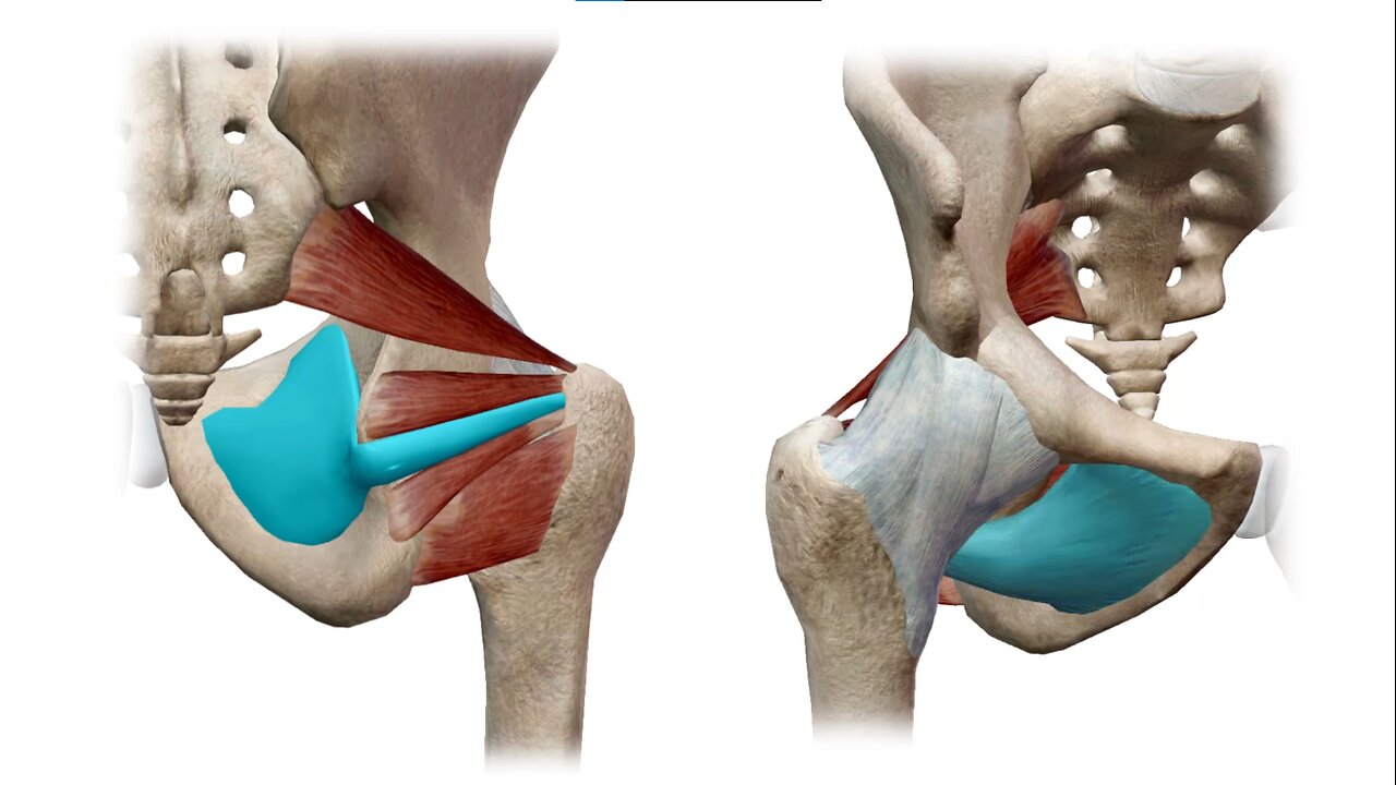
The labrum is a fibro cartilaginous rim attached to the margin of the acetabulum. It is horseshoe shaped and the ends are connected at the [00:01:00] acetabular notch via the transverse ligament. To form a complete circle, the labrum deepens the acetabular cavity to enhance joint stability. The labrum also creates a seal that helps keep synovial fluid within the articular cartilage, preventing direct contact between the femoral head and the acetabulum.
The labrum is wider and thinner in the anterior region of the acetabulum and thicker in the posterior region. There are several ligaments that connect the femoral head to the acetabulum to help provide further stability to the hip joint and restrict the joint from going into extreme positions. The most notable ligaments in the hip joint are the ilio femoral ligament, the pub femoral ligament, the ichi femoral ligament, and the ligamentum Terris.
The ilio femoral ligament is the strongest hip ligament and connects the pelvis to the femur at the front of the joint. This ligament is y shaped and often referred to as the Y ligament of Bigelow. It attaches to the anterior inferior iliac spine [00:02:00] and the lower part of the inter troon arteric line. It prevents hyperextension of the hip joint during standing by holding the femoral head within the aceta.
The is femoral ligament reinforces the capsule posteriorly. It originates on the isga part of the acetabular rim and spirals super laterally to the neck of the femur. This ligament also prevents hyperextension and holds the femoral head within the acetabulum. The Puba femoral ligament is attached to the Obterator crest and the superior Remus of the pubis and blends with the joint capsule.
The Puba femoral ligament reinforces the capsule anteriorly and inferiorly, and prevents over abduction of the hip joint. Now let's talk about the musculature of the hip. There are 22 muscles acting on the hip that contribute both to the stability and mobility at the hip. The musculature can be broken down into four quadrants anterior, posterior.
Medial and lateral. The anterior quadrant consists of the hip flexors, the ilio,
-
 1:21:30
Game On!
9 hours ago $1.26 earnedPatrick Mahomes does it AGAIN!
7.83K1 -
 15:20
15:20
Misha Petrov
2 hours agoReacting To Liberal MELTDOWNS Over Trump’s Return - Gen Z Is Planning a REVOLUTION?!
14.3K10 -
 18:22
18:22
Neil McCoy-Ward
2 hours agoThe 🇨🇴 Colombian President Just Learned The HARD WAY!!! (Another Win For The USA 🇺🇸)
13.3K7 -
 1:56:04
TheDozenPodcast
17 hours agoMass deportations, Islamists, Saving the UK: Nick Tenconi
25K18 -
 35:24
35:24
Survive History
22 hours ago $9.10 earnedCould You Survive in a Cavalry Regiment During the English Civil War?
81.1K8 -
 28:15
28:15
Degenerate Plays
16 hours ago $4.48 earnedTwo Birds' Secret Meeting - Gotham Knights : Part 26
64.3K5 -
 12:29
12:29
Mr. Build It
5 days agoWish I Knew This Before I Started Building It
69.1K25 -
 2:03:57
Megyn Kelly
2 days agoNew Trump Derangement Syndrome, and How CNN Smeared a Navy Veteran, w/ Piers Morgan & Zachary Young
163K188 -
 10:05
10:05
DIY Wife
3 years agoHow We Flip Old Furniture For Profit!
84.3K63 -
 2:14:54
TheSaltyCracker
14 hours agoTrump Goes Gangster ReeEEeE Stream 01-26-25
223K370