Premium Only Content

Gammaherpesvirus Infection of Human Neuronal Cells
Gammaherpesvirus Infection of Human Neuronal Cells
Importance: To date, no in vitro study has demonstrated gammaherpesvirus infection of neuronal cells. Moreover, worldwide clinical findings have linked EBV to neuronal pathologies, including multiple sclerosis, primary central nervous system lymphoma, and Alzheimer's disease. In this study, for the first time, we have successfully demonstrated the in vitro infection of Sh-Sy5y and Ntera2 cells, as well as human primary neurons. We have also determined that the infection is predominately LYTIC. Additionally, we also report infection of neuronal cells by KSHV in vitro similar to that by EBV. These findings may open new avenues of consideration related to neuronal pathologies and infection with these viruses. Furthermore, their contribution to chronic infection linked to neuronal disease will provide new clues to potential new therapies.
https://pubmed.ncbi.nlm.nih.gov/26628726/
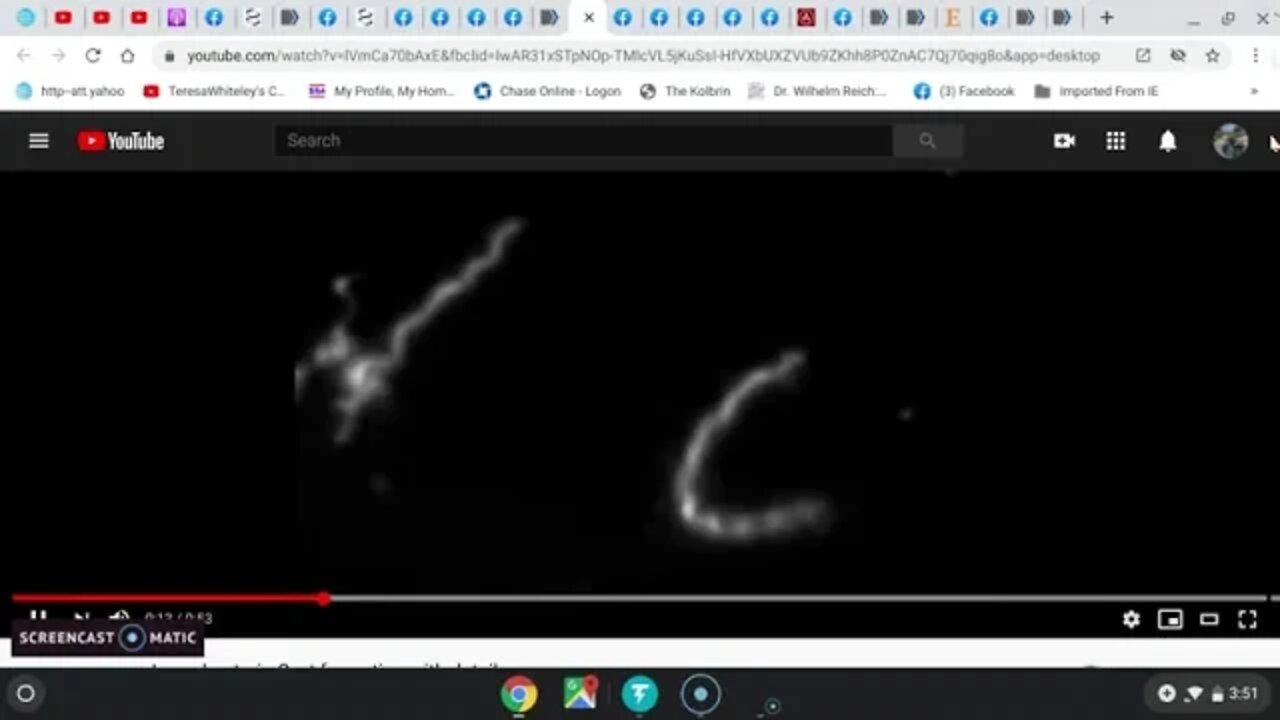
There's no such things as viruses. They are "lytic" ~bacterial associated, stress induced morphological forms of infinite antigenic variations in less than 2 seconds under antibiotic stress!
https://www.youtube.com/watch?v=lVmCa70bAxE&fbclid=IwAR11L3tmFlz8sIHO60hYned80en5X7BmQAQWwN7S4qOI3VTRWWpIdeuK2UE&app=desktop
https://pubmed.ncbi.nlm.nih.gov/27813134/
https://www.facebook.com/melthornburg0007/posts/10212167064805988
https://pubmed.ncbi.nlm.nih.gov/28935930/
Establishment and characterization of a novel Hodgkin lymphoma cell line, AM-HLH, carrying the Epstein-Barr virus genome integrated into the host chromosome https://pubmed.ncbi.nlm.nih.gov/27813134/
EBV infection is prevalent in the adenoid and palatine tonsils in adults
https://pubmed.ncbi.nlm.nih.gov/27864888/
Novel Spiroplasma Spp. Cultured From Brains and Lymph Nodes From Ruminants Affected With Transmissible Spongiform Encephalopathy
https://pubmed.ncbi.nlm.nih.gov/29155968/
Schizophrenia is Associated With an Aberrant Immune Response to Epstein-Barr Virus
https://pubmed.ncbi.nlm.nih.gov/30462333/
Morphological investigation of neurofibrillary tangles in Alzheimer's disease
https://pubmed.ncbi.nlm.nih.gov/7933715/
In it's molecular forms called Prion Proteins
In it's most basic forms it is called Myco PLASMA, Ana PLASMOSIS, Toxo PLASMOSIS or Spiro PLASMA--Exosomes, Blebs, Gemma, TAU. And your baaad cholesterol.
In it's viral forms it is host gene dependent LEGIONS.
In its bacterial forms it is MANY esp. Ehrlichia and H.pylori, TB, According to where it resides .
It's infectious organisms protected in fungus. You can kill the infections but the immune suppression they cause isn't easily fixed.
All of your so called viruses are simply morphological forms of infinite host gene variations.....Their cystic viral morphologies and secreted blebs containing seed formerly known as Prion and flagellum that recombines and grows with infectious DNA.
Discussion
The results reported here point to a possible primary target for the cytotoxicity expressed by the RepA-WH1 prionoid in E. coli: the bacterial internal cell membrane. Three distinct mechanisms have been proposed for membrane disruption by amyloidogenic proteins8, including: i) the formation of pores by the assembly of protein monomers into ring-shaped oligomers; ii) membrane thinning and iii) lipid (detergent-like) extraction. Since the last mechanism should imply the lysis of a significant fraction of the vesicles, which was not the case under our experimental conditions, and the second one seems incompatible with the observed persistence of aggregates bound to the membranes (Figs 3–5), the formation of pores by protein insertion into the lipid bilayer, as first proposed for Aβ
and α-synuclein39, was the most likely mechanism for vesicle leakage induced by RepA-WH1.
....leading to the characterization of RepA-WH1 as the first synthetic bacterial prionoid!
https://www.ncbi.nlm.nih.gov/pmc/articles/PMC4794723/pdf/srep23144.pdf
-

Epstein Borreliosis AIDS
2 years agoOPERATION WARPED SPEEDILY ROLLING OUT THE GREAT RESET OF HUMANITY AND THE BIOSPHERE
3462 -
 2:59:26
Tundra Gaming Live
8 hours ago $2.11 earnedThe Worlds Okayest War Thunder Stream//FORMER F-16 MAINTAINER//77th FS//#rumblefam
17K -
 2:32:19
DemolitionDx
4 hours agoSunday night COD with friends.
45.2K1 -
 2:10:14
vivafrei
14 hours agoEp. 237: More Trump Cabinet Picks! MAHA or Slap in the Face? Canada on Fire! Go Woke Go Broke & MORE
185K241 -
 2:23:21
SOLTEKGG
5 hours ago $3.20 earned🟢 First Day on RUMBLE!
37.3K3 -
 LIVE
LIVE
Vigilant News Network
8 hours agoCOVID-Vaccinated Hit With Grave New Reality | Media Blackout
1,843 watching -
 1:26:31
Josh Pate's College Football Show
8 hours ago $2.52 earnedSEC Disaster Saturday | Major CFP Earthquake Coming | Officiating Is A Disaster | New Studio Debut
25.8K2 -
 1:43:05
Adam Does Movies
11 hours ago $4.24 earnedGladiator II Spoiler Conversation With Hack The Movies
27.4K1 -
 24:10
24:10
Bwian
12 hours agoI Don't Know What I'm Doing in Fortnite, But I Still Won...
21.5K1 -
 19:30
19:30
DeVory Darkins
14 hours ago $41.46 earnedJoe Rogan MOCKS The View as Bill Maher HUMILIATES Woke Scientist
96.9K137